Scanning Probe Microscopy
Our atomic force microscopes are versatile tools for studying and characterizing surfaces or surface related properties. Since an AFM offers a multitude of operation modes not only the surface topography can be detected but also the spatial arrangement of electric and magnetic moments as well as differences in conductance, work function or friction, only to mention a few, can be visualized with highest resolution.
Basic Principal of Contact Mode

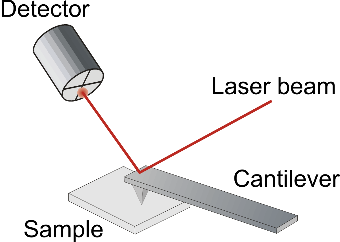
In an AFM a sharp tip at the end of a cantilever is brought in contact with the surface of a sample. Repulsive interatomic interactions between tip and sample lead to a deflection of the cantilever, which is read out with help of a focused laser beam and a segmented photodiode. A feedback loop controls the z-position of the tip by a piezo element in order to keep the deflection constant and ensure a stable contact between tip and sample. If we now scan the tip across the sample surface, the signal of the feedback loop gives us directly the information about the surface topography.
Imaging Ferroelectric Domains – Piezoresponse Force Microscopy

Piezoresponse Force Microscopy (PFM) is an ideal tool for the noninvasive imaging of ferroelectric domains on the micro-to-nano scale. Here we take advantage of the piezoelectric properties, the change of length due to the presence of an electric field, that is exists in any ferroelectric material.
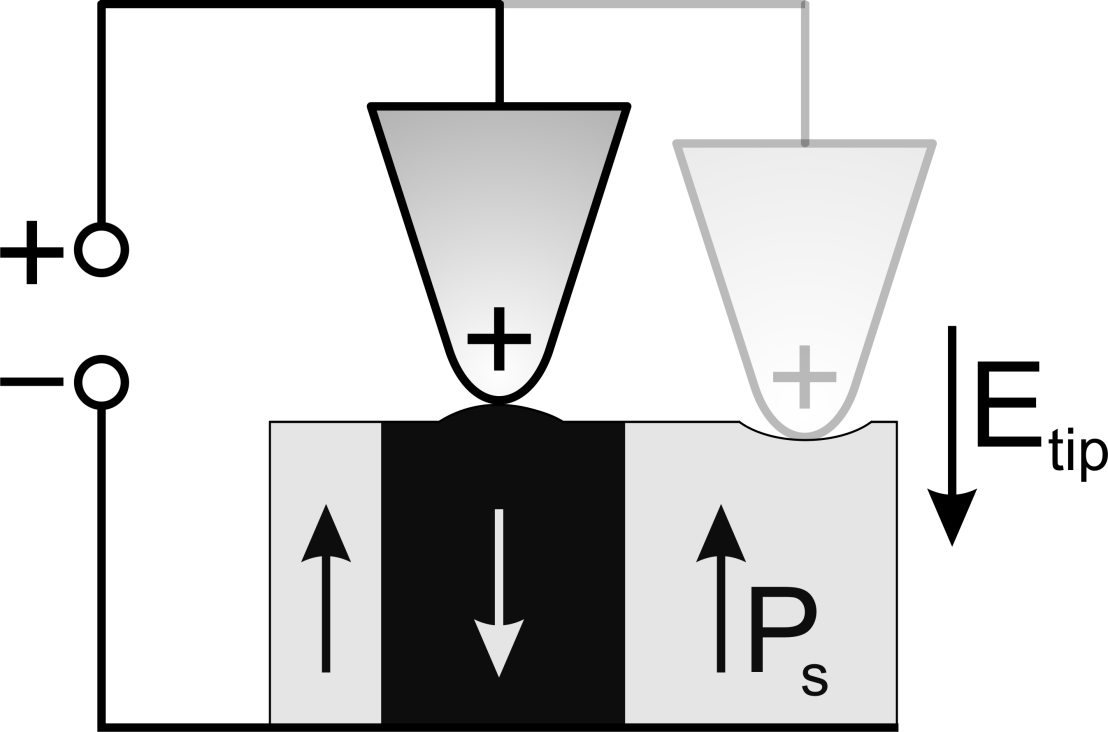
A small AC-voltage in the kHz regime is applied to the tip while the sample is grounded. The inhomogeneous electric field below the tip leads to the piezoresponse, a local deformation due to the piezoelectric effect, leading to a tiny deflection of the cantilever, which is detected by lock-in amplifier. For a certain field orientation the piezoresponse changes sign when crossing the boundary between two oppositely polarized domains, which leads to the observed image contrast.
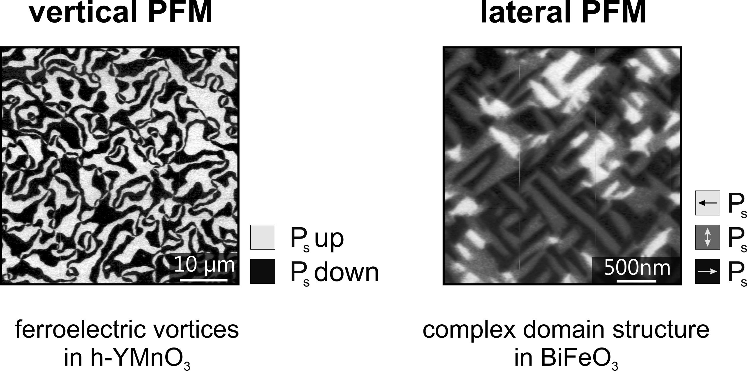
Since the displacement of the tip by the piezoresponse is not restricted to be directed parallel but also perpendicular to the surface normal, we distinguish between vertical and lateral PFM, which allows us to achieve information about the three dimensional orientation of the polarization.
Writing Ferroelectric Domains – Domain Engineering
Our PFM setup not only allows us to image ferroelectric domains with highest signal-to-noise level and high spatial resolution but also to manipulate and create ferroelectric domains by biasing the tip with a high DC voltage. The strong and inhomogeneous field below the tip reverses the direction of the spontaneous polarization. By controlling the position and bias of the tip we are able to write any domain structure with nm precision.

